The study of internal organs using ultrasound is considered one of the main diagnostic methods in various
fields of medicine. In cardiology, ultrasound of the heart, better known as echocardiography, which allows you to identify morphological and functional changes in the work of the heart, anomalies and irregularities in the valve apparatus.
Echocardiography (Echo KG) - refers to non-invasive diagnostic methods, which is very informative, safety and is carried out for people of different age categories, including newborns and pregnant women. This method of examination does not require special training and can be carried out at any convenient time..
Unlike X-ray examination, (Echo KG) can be carried out several times. It is completely safe and allows the attending physician to monitor the patient's health and the dynamics of cardiac pathologies. During the examination, a special gel is used, which allows ultrasound to better penetrate the heart muscles and other structures.
What allows you to examine (ЭхоКГ)
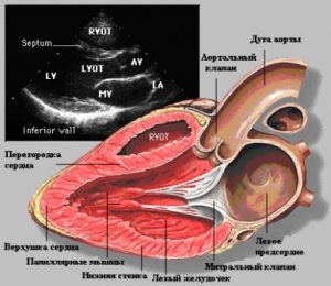
Ultrasound of the heart allows the doctor to determine many parameters, norms and deviations in the work of the cardiovascular system, estimate the size of the heart, cardiac cavity volume, wall thickness, beat frequency, presence or absence of blood clots and scars.
Also, this examination shows the state of the myocardium., pericardium, large vessels, mitral valve, the size and thickness of the walls of the ventricles, determines the state of the valve structures and other parameters of the heart muscle.
After the (Echo KG) the doctor records the results of the examination in a special protocol, decoding of which allows you to detect cardiac diseases, deviations from the norm, anomalies, pathology, also diagnose and prescribe appropriate treatment.
When to do (Echo KG)
recurrent or frequent heart pain;
rhythm disturbances: arrhythmia, tachycardia;
dyspnea;
increased blood pressure;
signs of heart failure;
previous myocardial infarction;
if there is a history of heart disease;
It is possible to undergo this examination not only in the direction of a cardiologist, but also other doctors: endocrinologist, gynecologist, neurologist, pulmonologist.


 The study of internal organs using ultrasound is considered one of the main diagnostic methods in various